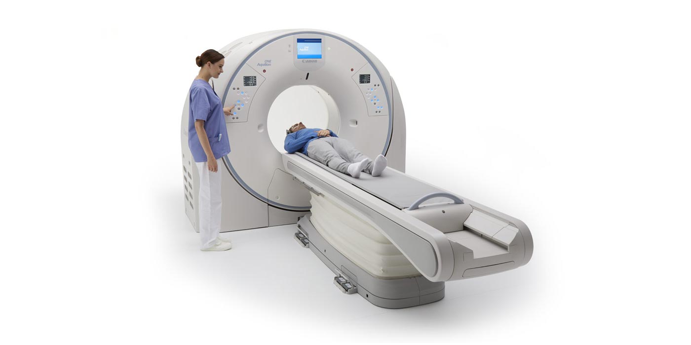
A CT scan uses specialized x-ray beams to create highly-detailed pictures of the body. The special x-ray beams pass cross-sectionally through the body and are received by a special detector array, which separates the beam into very thin slices. The high-resolution slices are typically grouped into volumes of data which can be reconstructed into any desired plane without losing resolution (clarity).

Magnetic resonance imaging (MRI) is a method of looking inside the body using magnetism and radio waves to produce remarkably clear pictures of the body, such as brain, spine, and joints. An MRI scanner consists of a strong magnet with a radio transmitter and receiver. MRI produces soft-tissue images and is used to distinguish normal, healthy soft tissue from pathologic (diseased) tissue.
Meenakshi Group of diagnostic centers utilizes a 1.5 Tesla (1.5T) MRI system which refers to the field strength of the magnet. 1.5T MRI provides the highest quality, most accurate diagnostic images. There are many small structures in the body and it is important for clinician to be able to see them with as much detail as possible.
MRI is commonly used to look at the brain, spine and nerves, joints such as shoulders and knees, blood vessels such as the carotid arteries, breasts, and abdominal organs such as the liver.
In some circumstances, a contrast agent (gadolinium) is injected to give the radiologist more information about a specific body part as it looks at the blood flow to a particular area of interest.
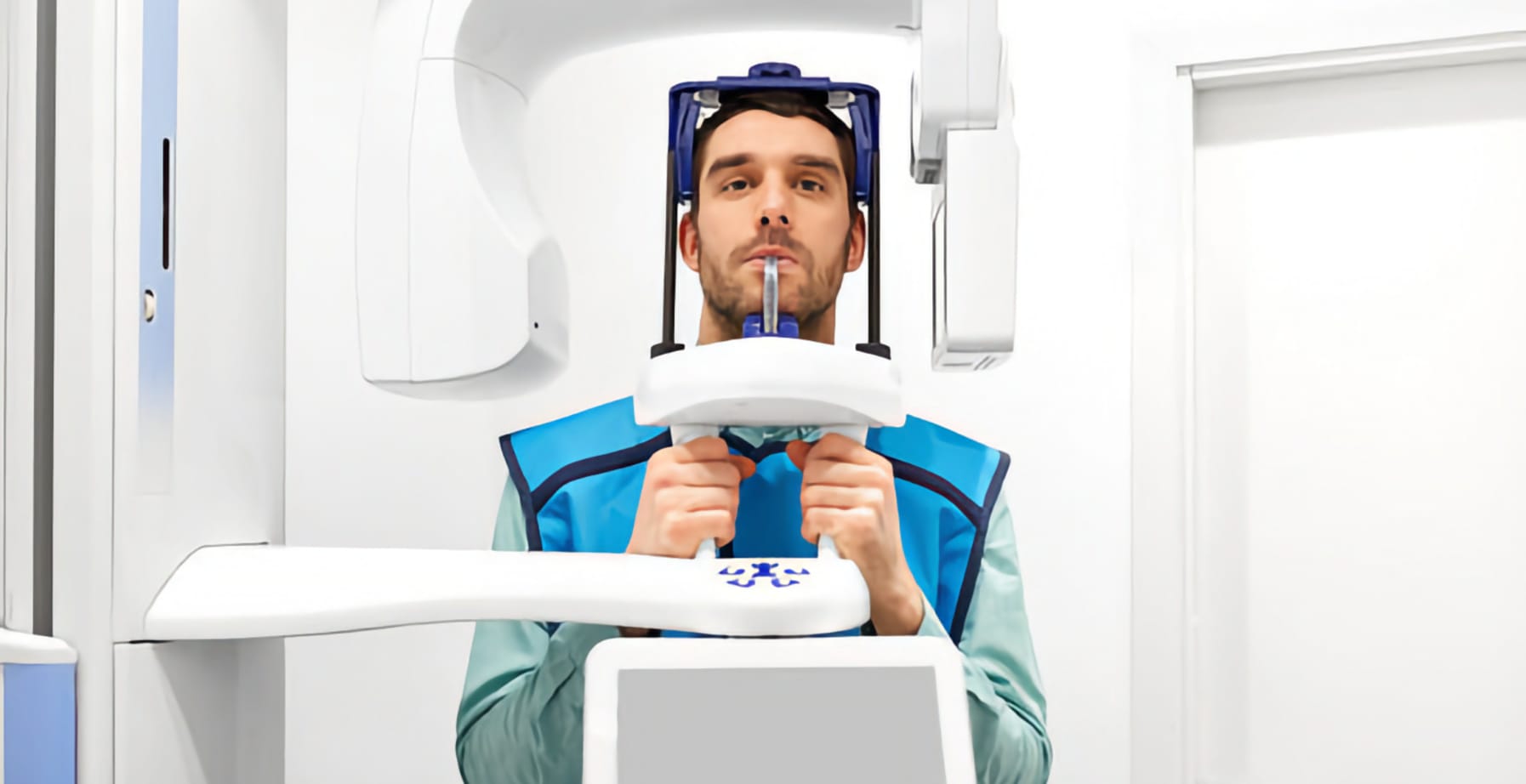
An OPG also called an Orthopantomogram, is a panoramic scanning dental X-ray. It provides a broad view of the mouth, teeth, and bones of the upper and the lower jaws. With an OPG X-Ray, your doctor can see all the teeth numbers, positions, and growth. The dentist can also see the teeth which have not yet erupted through the gum. Meenakshi Group of Diagnostic Centre’s offers Radiological a Services to all. An OPG in Meenakshi Group of Diagnostic Centre’s also shows issues with the jawbones and the joint that connects the mandible to the head called TMJ. TMJ refers to Temporomandibular joint.
An OPG X-Ray shows a flattened two-dimensional view of a half-circle, i.e., from ear to ear. This takes images from multiple angles to make up the compounded wide image, where the maxilla (upper jaw) and mandible (lower jaw) are in the viewed area. The structures present outside the viewed area are blurred in the image. The OPG X-Rays are different from the small close-up X-Rays. Small close-up X-Rays are used to view individual teeth, whereas an OPG covers a much broader area. OPG can go to hard-to-see spaces like wisdom teeth, and the development of a child’s jaw and teeth. It also checks the jaw joint, the Temperomandibular joint (TMJ).
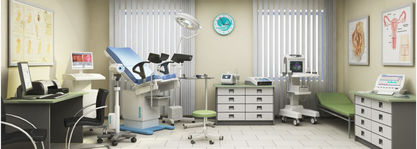
Ultrasound Scan (Sonography) is a procedure that uses high-frequency sound waves to build live images. Ultrasound scans, or sonography, are preferred as they use sound waves to produce an image alternately of radiation. An Ultrasound is widely used during pregnancy. In Meenakshi Group of Diagnostic Centres Ultrasound scan are practiced to appraise fetal development, and they can distinguish problems in the liver, heart, kidney, or abdomen. They may furthermore serve in achieving certain types of biopsy.
The image that dawned is called a sonogram.
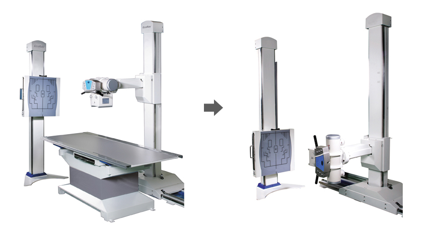
X-Rays use a type of radiation called electromagnetic waves to produce images of structures in our body like joints and bones. Meenakshi Group of Diagnostic Centres offers best X-Ray. It is a painless method used to display images inside of the body and is carried by trained specialists called radiographers.
X-Ray is a commonly used way to detect a range of abnormalities inside the body in different shades of black and white. Dense materials, such as bones are displayed as white on X-rays, the air in the lungs shows up as black and the fat and muscle appear in gray shades. This is because some tissues receive different quantities of radiation. , Meenakshi Group of Diagnostic Centres a leading diagnostic centre offers best X-Ray services under the guidance of expert Radiologist
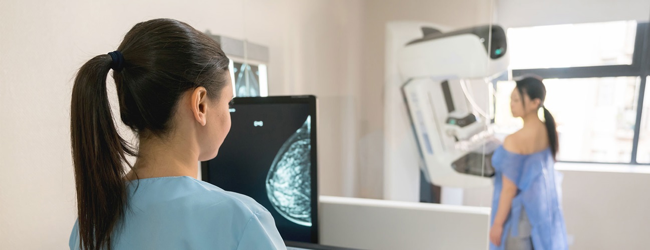
Mammography is a method of x-ray imaging used to diagnose breast cancer in women before women experience symptoms. It is the most common way to diagnose breast cancer, and the information provided in mammography can save lives. This tests help to reduce the number of deaths from breast cancer in women of age 40s to 70s. Mammography in Meenakshi Group of Diagnostic Centres may also show few other irregularities present in the breast and breast problems like pain, lumps and nipple discharge.
Mammography device consists of two firm surfaces. Women stand in front of an X-Ray machine during the Mammography process, and the breast is placed between the two surfaces. These surfaces compress and spread the breast, and images are captured from two different angles. The images captured in Black and white color will be displayed on the monitor. Women feel a little uncomfortable during the process as the breast gets flattened by applying some pressure for a few moments. Meenakshi Group of Diagnostic Centres offers best Mammography scan.
Our Services
At Meenakshi Group of Diagnostic Centers we are providing world class Diagnostic services under one roof with accuracy and precision.

MRI
Method of looking inside the body using magnetism and radio waves to produce remarkably clear pictures of the body, such as brain, spine, and joints.
View More
CT Scan
A CT scan uses specialized x-ray beams to create highly-detailed pictures of the body. The special x-ray beams pass cross-sectionally through the body and are received
View More
Orthopantomogram
An OPG also called an Orthopantomogram, is a panoramic scanning dental X-ray. It provides a broad view of the mouth, teeth, and bones of the upper
View More
Mammography
Mammography is a method of x-ray imaging used to diagnose breast cancer in women before women experience symptoms. It is the most common way to diagnose breast cancer
View More
PET
Positron Emission Tomography (PET) is a powerful imaging modality widely used in nuclear medicine for diagnosing and monitoring a variety of conditions,
View More
Gamma
The Gamma Camera, also known as the Scintillation Camera or Anger Camera, is a fundamental imaging device in nuclear medicine. It operates on the principle of
View More
X-Rays
X-Rays use a type of radiation called electromagnetic waves to produce images of structures in our body like joints and bones. Meenakshi Group of Diagnostic
View More